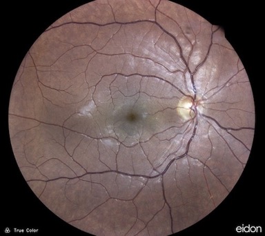
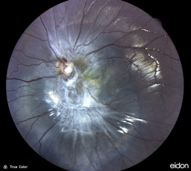
Acute Macular NeuroRetinopathy (AMNR) COVID, Combined Hamartoma of Retina and Retinal Pigment Epithelium
Update January 29, 2022
New Icare Eidon UWFL
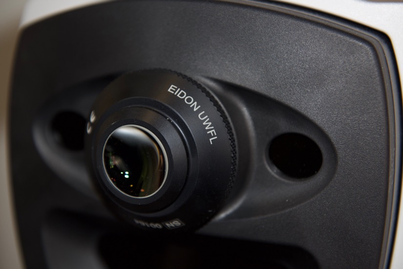
Eidon /New Eidon UWFL
click to enlarge (and then close window)
Diabetes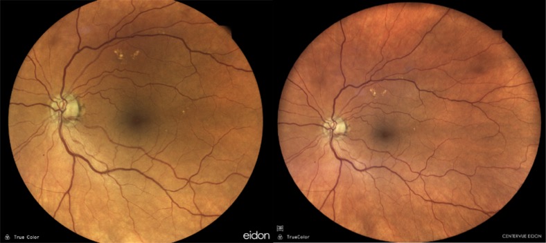
Macular hemorrhage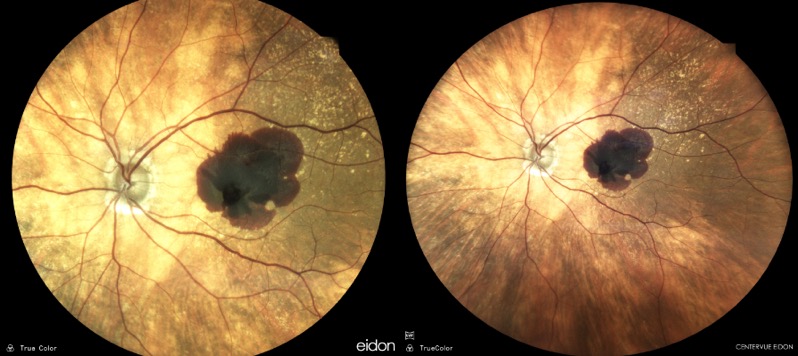
Toxo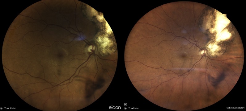
And my beautiful fundus: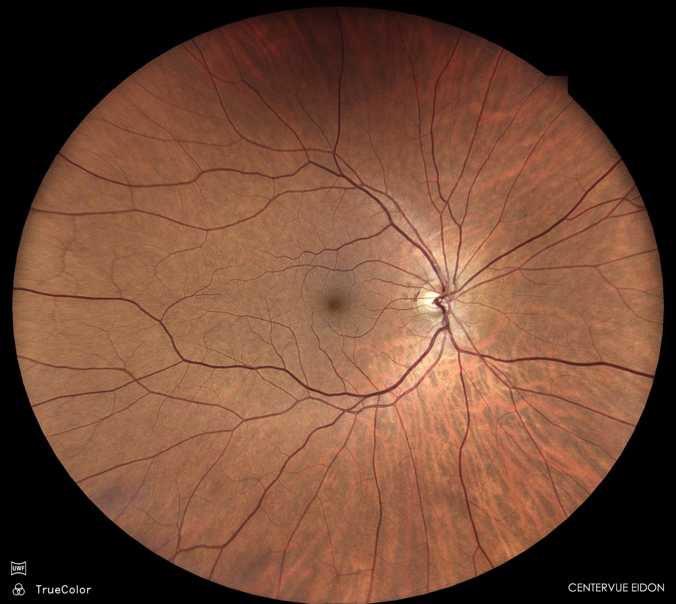
Update September 26, 2020
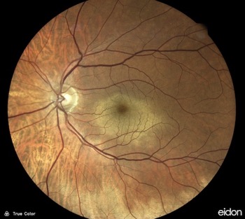
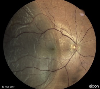
Macular hemorrhage (Valsalva retinopathy), Bungee cord trauma, Retinal detachment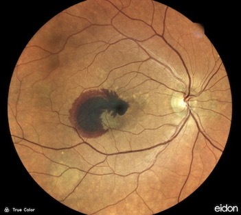
Update July 2, 2020
Update June 10, 2020
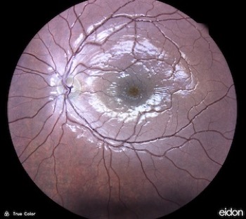
X-linked retinoschisis, arterial occlusion, branch retinal vein occlusion, retinal vein occlusion and retinal detachment
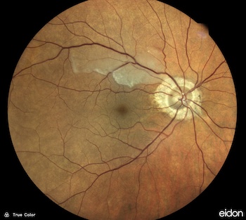
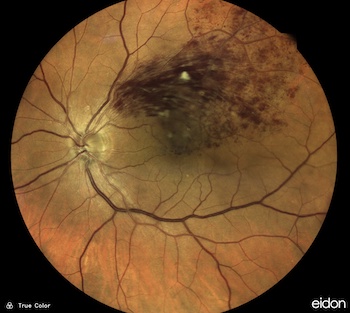
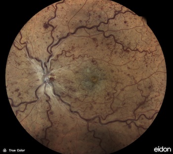
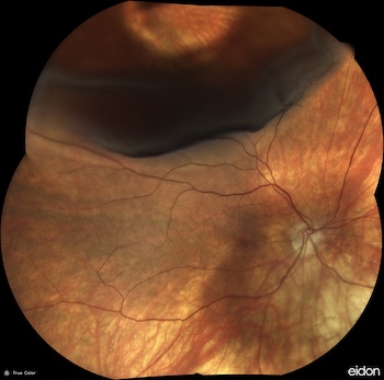
Update January 04, 2020
a fantastic meeting: SINGAPORE
1st Asia-Pacific Ocular Imaging Society Meeting / 17-19 january, 2020 APOIS
Dr Jean-Michel Muratet, Fabian Chatwin Cedrati (Centervue) and Joonas Ihalainen (Icare)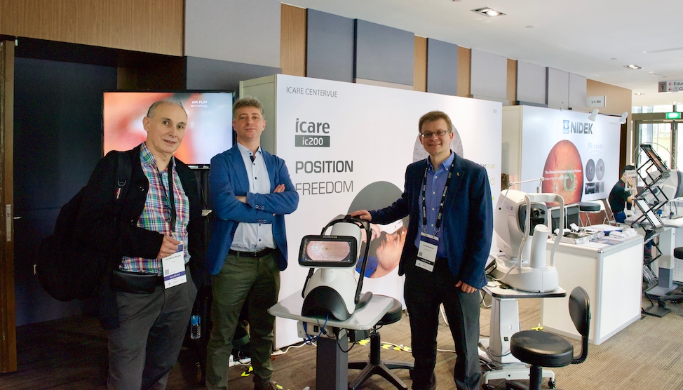

Update May 17, 2019
Société Française d'Ophtalmologie PARIS, May 11-14, 2019, chorioretinal folds (posterior scleritis), and retinitis pigmentosa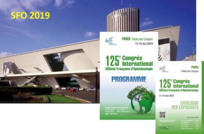
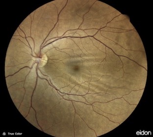
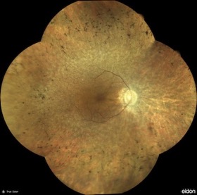
Update April 20, 2019
Japanese Ophthalmological Society (April 2019), venous and artery occlusion, retinal embolism, OCT angiography congress (ROME) December 13-14, 2019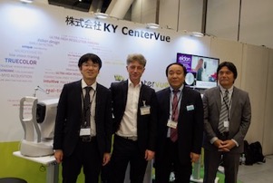
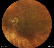
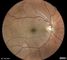
Update January 14, 2019
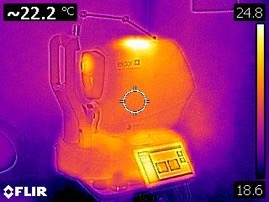
Japanese Ophthalmological Society (April 2019), Eidon thermographic imaging
Update December 08, 2018
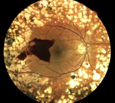
Rome, diabetes, diabetes, retinal detachment, Toulouse January and direct blow to the eye in tennis 
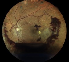
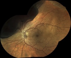
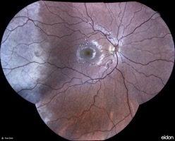
Update October 12, 2018
Fluorescein Angiography Eidon (FA Eidon) CenterVue Italy, American Academy of Ophthalmology 2018 (Chicago) and artery occlusion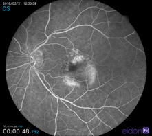

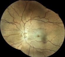
Update July 06, 2018
X-linked retinoschisis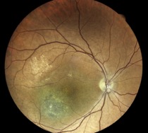

Update May 19, 2018
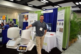
Arterial occlusion, and ARVO 2018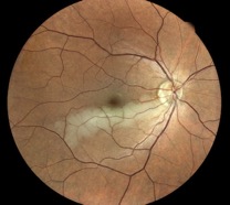
Update February 12, 2018
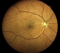
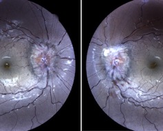
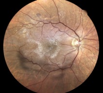
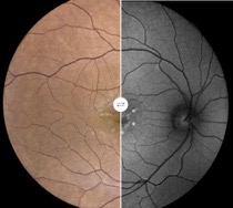
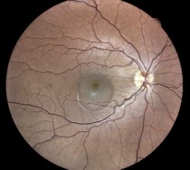
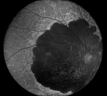
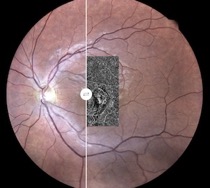
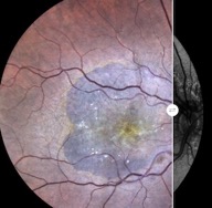
Retinitis pigmentosa, artery occlusion, papilledema, epiretinal membrane, Stargardt disease, Central Serous Chorioretinopathy (CSC), another Stargardt disease and traumatic choroidal rupture 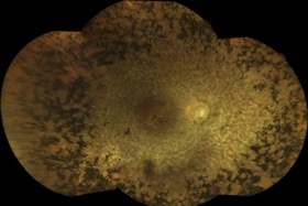
Update December 10, 2017
Hong-Kong Meeting with Fabian Cedrati (CenterVue-Italy) and Eye doctors
Thank you Fabian !!
Credit photos http://www.muratetphotographie.com
Update October 8, 2017
Atelier OCT-Angiography Dr Florence Coscas October 20 (Paris France), and a video about Eidon & OCT Optovue 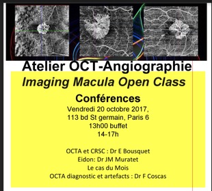
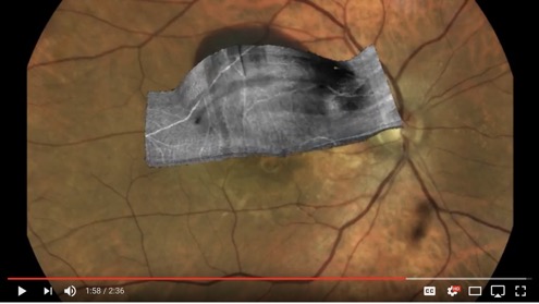
Update: october 08, 2017
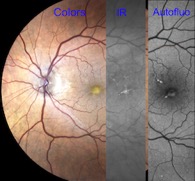
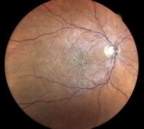
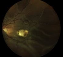
Eidon + OCT 3D (Optovue) RPE tears (ARMD), diabetes, vitelliform, epiretinal mb, retinal detachment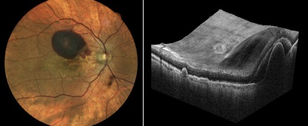
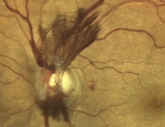
Update: July 27, 2017
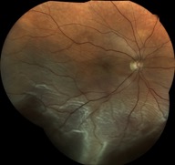
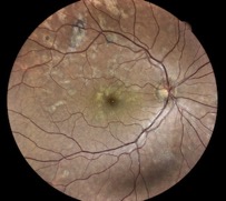
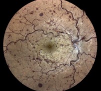
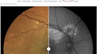
Eidon + OCT angiography AngioVue Optovue (video), retinal detachment, APMPPE, retinal venous occlusion, chronic serous chorioretinopathy and Retinal artery occlusion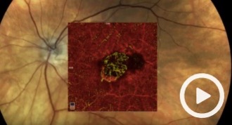
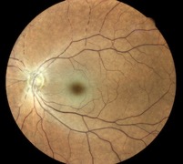
[Update: June 13, 2017]
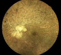
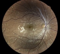
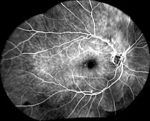
Chloroquine maculopathy, retinitis pigmentosa, diabetic retinopathy and fluorescein angiography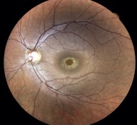
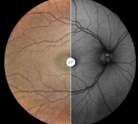
[Update: June 01, 2017]
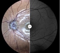
American EIDON ARVO team (bottom of this page), autofluorescence of papillary drusen (with Angioid Streaks and Pseudoxanthoma Elasticum), ocular albinism, AMD (with OCTA)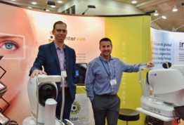
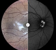
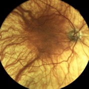
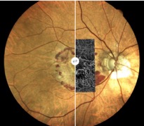
[Update: April 28, 2017]
Two composite AMD images (Eidon image + OCT angiography AngioVue OPTOVUE), 3 toxoplasmosis and one combined hamartoma of retina and retinal pigment epithelium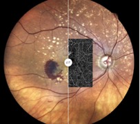
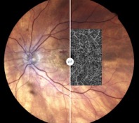

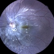
[Update: April 12, 2017]
New autofluorescence image (AMD), boxing, metastasis of breast carcinoma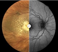
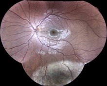
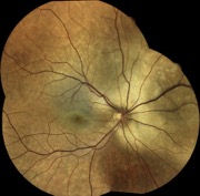
[Update: Februrary 2, 2017]
Le rétinographe EIDON de la société CenterVue permet une visualisation exceptionnelle de la rétine, grâce à une technique spécifique, il devient "The first True Color Confocal Scanner".
La visualisation du fond d'oeil devient évidente, malgré la présence d'opacités des milieux (cataracte ou hémorragie du vitré).
La prise de vue est possible avec de petites pupilles (jusqu'à 2,5mm)
Cet appareil est un lointain cousin des rétinographes non-mydriatiques (RNM) habituels, et la comparaison de leurs images nous permet de percevoir très rapidement les différences.
L'option autofluorescence dans le bleu ajoute des capacités impressionnantes à cet appareil qui va devenir indispensable dans bon nombre de cabinets d'ophtalmologie.
Je suis content de partager avec vous l'explosion des technologies médicales de pointe et leur mise à disposition.
J'ai vécu avec satisfaction la diffusion mondiale des OCT (Optical Coherence Tomography) en 2008, et la possibilité d'utiliser un EIDON aujourd'hui complète ainsi l'exploration multimodale de la rétine, au bénéfice de tous nos patients.
Je remercie tous les confrères qui m'ont envoyé des images fantastiques, et je reste à la disposition de tous pour mieux comprendre les images que l'on acquiert.
Le 2 février 2017
Dr Jean-Michel Muratet
09100 Pamiers
France
No financial interest
Japanese Ophthalmological Society (April 2019)
Dr Jean-Michel Muratet & Fabian Cedrati (CenterVue)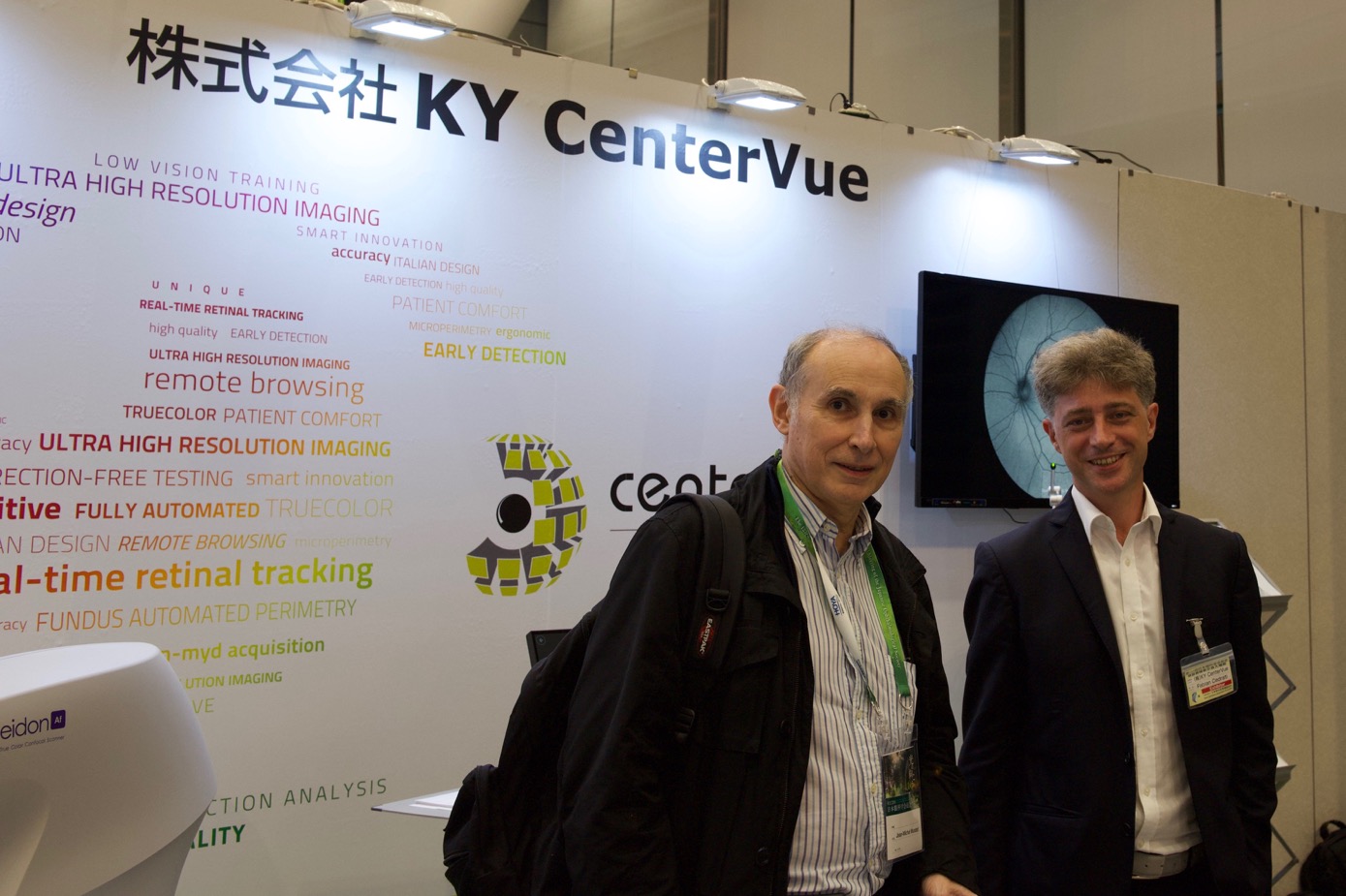
Le mot Eidon vient du grec ancien, et correspond à une conjugaison du verbe voir (orao = je vois ou ὁράω). Ce temps du passé qui n'existe pas en Français est l'aoriste et donne eidon (εἴδον), ce qui pourrait se traduire par "j'ai vu".
Une comparaison d'un RNM habituel et d'une rétinophotographie Eidon, avec un trouble du milieu (cataracte):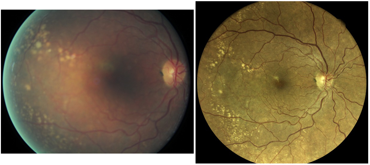
RNM vs EIDON Courtesy of Dr Vincent Gualino, Montauban, France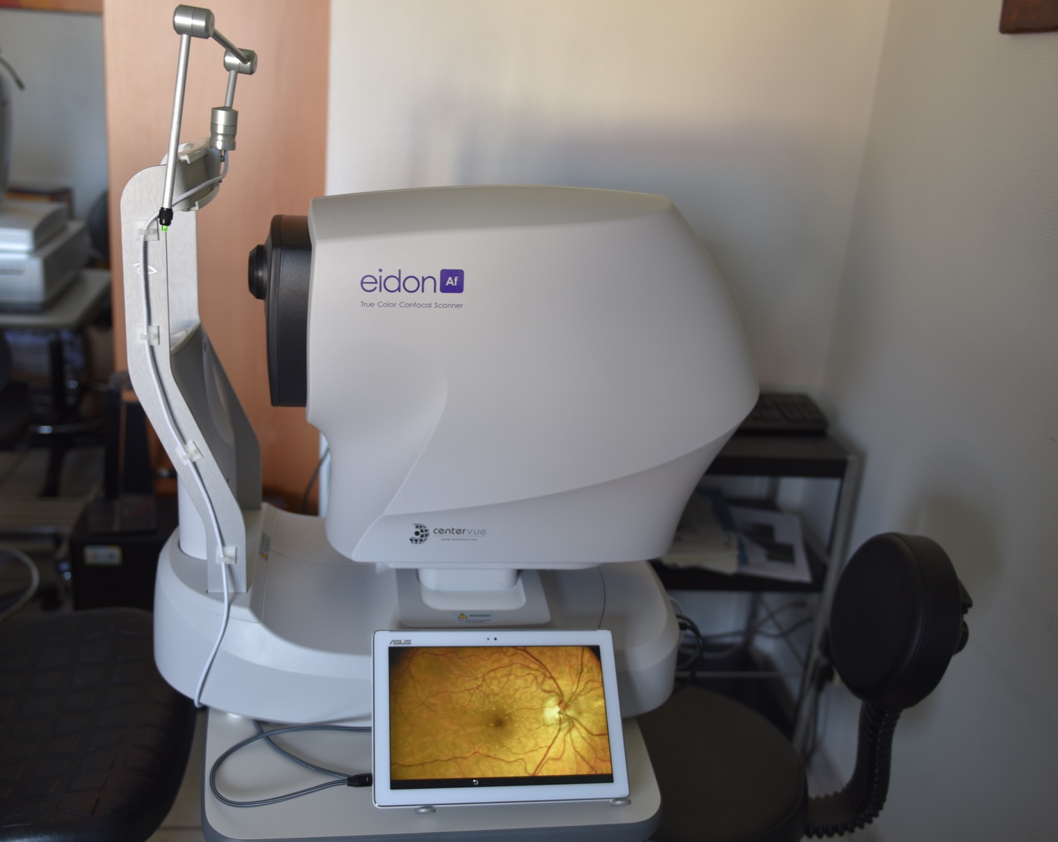
Courtesy of Dr Jean-Michel Muratet, Pamiers, France
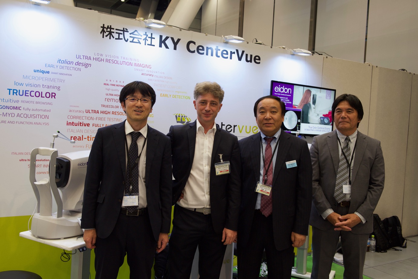
Japanese Ophthalmological Society CenterVue team (Tokyo, Japan, April 2019):
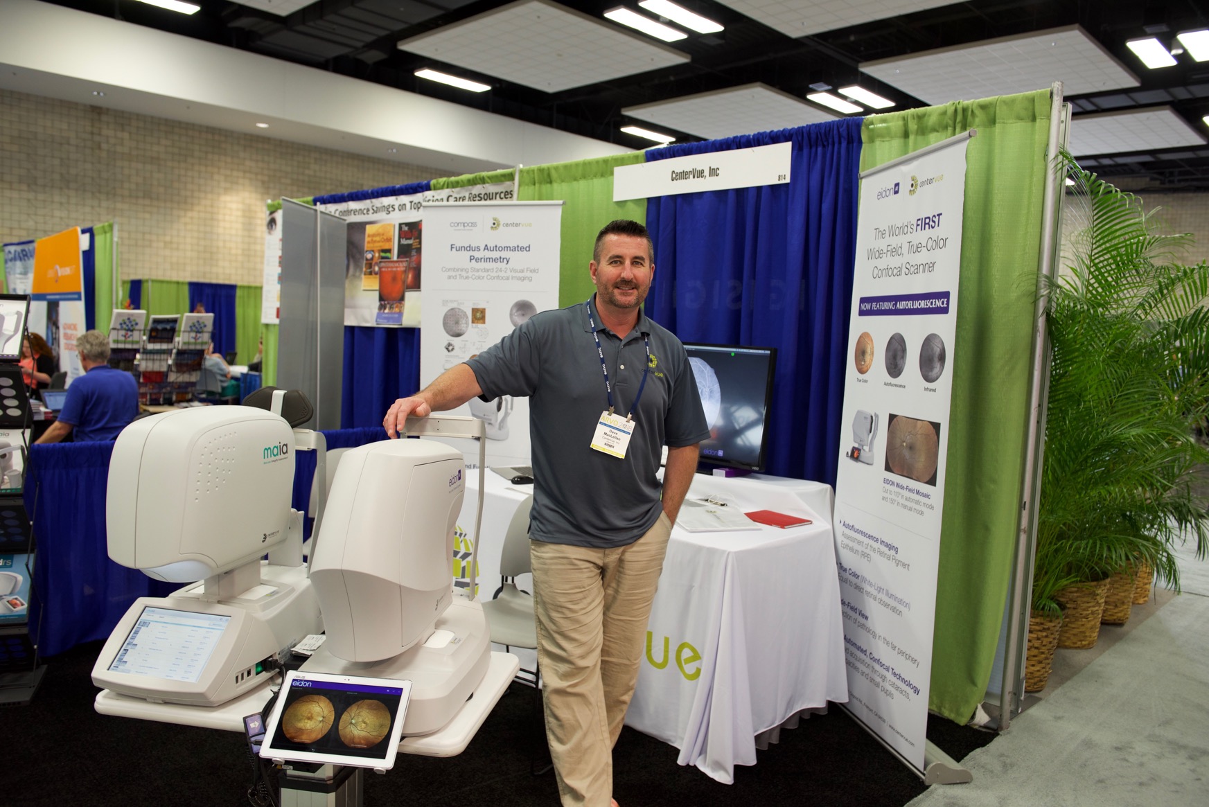
American EIDON ARVO team (Honolulu, USA, May 2018):
French EIDON dream team (JRO March 2017, Paris, France):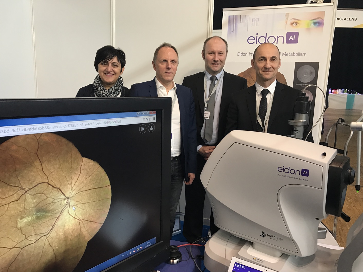
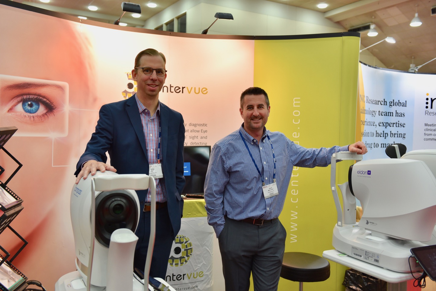
American EIDON ARVO team (Baltimore, USA, May 2017):
No financial interest
